Medical Image Analysis: Improving Patient Outcomes
Researchers
Professor Reyer Zwiggelaar
Dr Chuan Lu
Dr Yonghuai Liu
The Overview
Medical image processing allows for in-depth, but non-invasive exploration of internal anatomy. It has become one of the key tools leveraged for medical advancement in recent years. Research on medical image analysis within the Vision Graphics & Visualisation (VGV) research group at Aberystwyth University (AU) has led to a wide range of developments in healthcare informatics.
The Research
Research conducted by the VGV research group has led to a significant number of healthcare developments, particularly around commercial orthopaedics’ segmentation software, the International Deep Endometriosis Standard, Multiple Sclerosis (MS)/stroke segmentation and stroke rehabilitation, and retinal disease treatment. It has improved patient outcomes, from individual cases through to a group of hospitals. It has resulted in changing practices with newly introduced international standards in the relevant healthcare sector, and has benefitted the commercial sector with improved tools that in turn enhance patient outcomes.
The Impact
MRI/PET data has predominantly been used for breast/prostate research. The VGV research group extended the data use into the area of brain segmentation, with an emphasis on Multiple Sclerosis (MS) lesions. Concurrently, the group collaborated with the University of Girona, as a main contributor to two significant MS lesion-driven projects. These projects led to new pre-processing techniques, established by the VGV research group, and directly contributed to the creation of Tensormedical, a medical company who provides lesion analysis in clinical environments. This not only helps clinical experts but also contributes directly to patient well-being. In recognition of this research, the VGV research group has received significant grants to extend it to dealing with the rehabilitation problems of stroke patients from NHS Wales and Health and Care Research Wales.
The International Deep Endometriosis Analysis (IDEA) group statement is the first international consensus on nomenclature and measurements in endometriosis imaging. Methodologies developed by the VGV research group have been included as sonographic steps for endometriosis diagnosis, published as a consensus opinion by the International Deep Endometriosis Analysis Consensus Group, significantly impacting clinical practice in endometriosis. Methodologies include a preoperative Ultrasound-Based Endometriosis Staging System (UBESS), developed to predict the level of complexity of laparoscopic surgery for endometriosis, and the development and evaluation of Transvaginal Sonography (TVS) techniques for diagnosis and management of endometriosis.
The research has also provided underlying evidence for the National Institute for Health and Care Excellence (NICE) guideline on the standard of using ultrasound imaging as cost-effective tools for endometriosis.
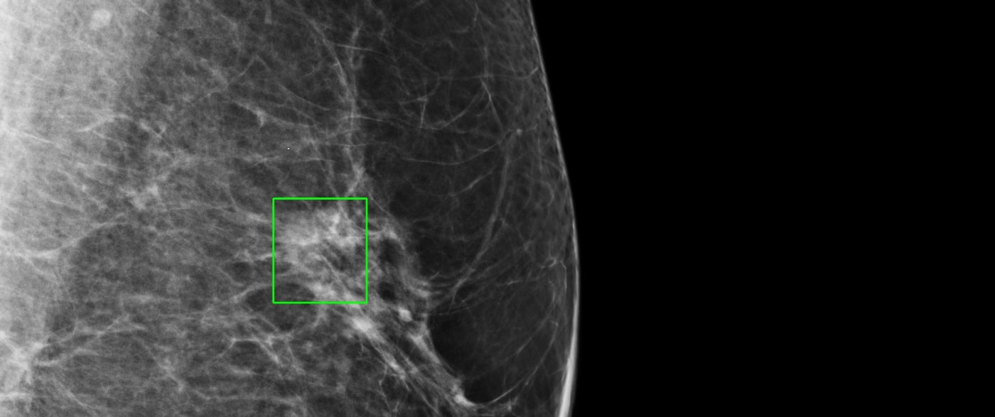
Research on segmentation techniques in medical image analysis has been used extensively in the development of computer-aided diagnosis (CAD) techniques. Over the years this has had a strong emphasis on mammographic and prostate-based applications, where the segmentation techniques formed the essential pre-processing step before further analysis/classification and clinical recommendation might be possible. Such pre-processing steps enable the establishment of novel mechanisms that can exploit texture and intensity topology natures of image data. This research has resulted in, and subsequently benefitted from, extensive academic collaboration with international research groups including the universities of Girona, Pennsylvania and Manchester Metropolitan. The main impact in this area has been the translation of mammographic image analysis techniques to a range of applications in the commercial sector. Typical examples for this are the orthopaedic segmentation tools developed at Synopsys, a major software company, and the CT Liver analysis software developed at Toshiba Medical Systems, which are being extensively used in the clinical domain. These commercial tools significantly contribute to clinical reporting and improving patient outcomes.
The original retinal work is based on Retinex research developed within the VGV research group. Through exploiting novel 2-D / 3-D symmetric filters, the process of detecting vascular and other structures is automated, improving understanding of the mechanism, diagnosis, and treatment of many vascular pathologies.
This research has been conducted in close collaboration with a group of clinical end-users tackling a range of retinal specific clinical challenges, which include lesion and vascular segmentation/classification.
The work on retinal images is directly based on an earlier investigation into linear structures in medical images. The incorporation of automated segmentation for the tortuosity of corneal nerve fibres has provided a consistent approach to the assessment of dry eye disease and diabetic neuropathy. At the same time, it has significantly accelerated the processing time of the staging of patients, increasing the volume of patients being treated by 10%. This has meant that for a single hospital (Peking University Third Hospital) 200 more patients can be assessed and treated each year, with similar effects in the Royal Liverpool University Hospital. This also led to a recent collaboration with Hywel Dda University Health Board where retinal scans can be linked to neurological and mental health diseases.
In recognition of their research, the VGV research group has received significant grants to support the rehabilitation problems of stroke patients from NHS Wales and Health and Care Research Wales.
Get in touch
As a University, we’re always keen to share our knowledge and expertise more widely for the benefit of society. If you’d like to find out more or explore how you can collaborate with our researchers, get in touch with our dedicated team of staff in the Department of Research, Business and Innovation. We’d love to hear from you. Just drop an e-mail to:
research@aber.ac.uk
Research Impact Case Studies | Research Theme: Health


